An international group of researchers are looking for owners of normal healthy horses as well as owners of horses that headshake to complete a short online survey to help understand headshaking. They are trying to understand why some horses develop headshaking behaviour and others do not. With a better understanding it is hoped that more effective treatments can be developed.
The group have launched a simple online survey and the project is funded by UK based Science Supplements Ltd. The group want to hear from owners who have horses that headshake BUT just as importantly, from owners whose horses don't headshake. These horses will act as controls for the study.
To date the group have over 300 completed surveys from owners of horses that headshake and from over 400 that don’t. The group urgently need more surveys, especially from owners of horses that DO NOT HEADSHAKE. (Please note that at this time the survey is limited to horses in the UK)
The survey takes around 10 minutes to complete and can be found here:
https://www.surveymonkey.com/s/HEADSHAKING2014

Thursday, May 08, 2014


Saturday, February 15, 2014
Is crib-biting inherited?

Various factors have been suggested as a cause for the behaviour including gastric discomfort and lack of sufficient environmental stimulation. But is there a genetic component?
Scientists at the University of Helsinki, Finland compared crib-biting and non-crib-biting horses, looking specifically at genes known or suspected to be related to stereotypic behaviours.
In particular they looked at genes such as Ghrelin, Ghrelin receptor, Leptin, Dopamine receptor, μ-opioid receptor, N-cadherin, Serotonin receptor and Semaphorin.
Two groups of horses were compared. Horses in the crib-biting group had started to display the behaviour at any early age, and had done so for at least a year. They tended to crib-bite after feeding or when stressed.
Non-crib-biting (control) horses were all over ten years old and had never been seen to crib-bite.
The researchers analysed the candidate genes in both groups and compared the allele frequencies between the cases and controls for each breed separately. They could find no evidence of an association at any of the tested loci.
They conclude: “These results suggest that the previously known stereotypic genes are not major risk factors for crib-biting in horses.” They suggest that further whole genome studies involving larger groups of crib-biting and non-crib-biting horses are required.
More information
Thursday, February 13, 2014
Detecting larval Strongylus vulgaris infections
Collaboration between scientists in Denmark and the United States has led to the development of a diagnostic test that promises to help identify horses infected with the larval stages of the large strongyle Strongylus vulgaris. The larvae migrate in the arteries that supply the large intestines and may cause blockage of the arteries and intestinal infarctions.
The result of the research is a serum enzyme-linked immunosorbent assay (ELISA) which has been validated for detecting migrating larvae in the bloodstream of horses. The test detects antibodies against a polypeptide in the excretory/secretory fraction of the migrating larvae, named SvSXP.
Further work using the test showed that foals receive colostral antibodies from the mare which may persist for up to 13 weeks of age. Foals start being able to mount their own immune response to the parasites from about three months of age, depending on the grazing conditions and the stage at which they were exposed to the parasites.
In younger foals, the serum ELISA does not give reliable diagnostic information despite heavy infections. The researchers found that young foals can be heavily infected without showing positive ELISA test results.
They also found that higher infection levels tend to be associated with higher ELISA levels.
Once they had started producing antibodies, foals continued to maintain high ELISA levels throughout the study. There was a tendency for the ELISA levels to decline gradually as the numbers of migrating larvae declined and adult intestinal worm burdens increased.
The scientists suggest that further research into the time taken for antibody levels to decline after effective anthelmintic treatment would be very useful.
If the test becomes commercially available it will be a useful addition to the armoury of diagnostic tests for investigating parasitic disease in horses.
Read more at equinescienceupdate.com
Monday, February 10, 2014
Investigating sudden death in racehorses

Recently Catriona Lyle led a multicentre study in an attempt to gain a better understanding of the problem and find ways to reduce the risk of sudden death occurring. The work, funded by a Horserace Betting Levy Board scholarship, was undertaken at the Royal (Dick) School of Veterinary Studies in Edinburgh.
Lyle coordinated the study drawing information from racecourses in North America, Australia, Japan and Hong Kong. Post-mortem data from 284 cases over 20 years were studied. In the UK post-mortem examinations are not always carried out in cases of sudden death and so gaining access to these international records was essential.
She explains:“The study has shown that the cause of death can be quite variable, ranging from severe lung bleeding to a pelvic fracture that causes massive bleeding into the abdomen. But in approximately half the cases I studied, the pathologist was uncertain as to the cause of death. The most likely explanation for death in these situations is cardiac rhythm irregularities, but this is very difficult to prove.”
Following her analysis of these international data, Lyle then looked at cases of sudden death in British racehorses. She found that sudden death of horses during races is a very rare event. Over a seven year period, in 705,914 race starts, there were 201 sudden deaths on British racecourses. The same syndrome is known to occur in eventing, show jumping and hunting but statistics have not been established in these sports.
In the UK study, she found that increasing age is a risk factor, steeplechases posed more of a risk than flat races and racing during the summer was associated with a greater risk of sudden death. However, this should be put in the context that, on average, steeplechasers are older than hurdlers or Flat horses. Horses that had raced within the last 60 days were less likely to be affected.
Read more at equinescienceupdate.com
Thursday, January 23, 2014
All you need to know about equine hormonal disorders?
Up
to date information on equine hormonal disorders has just become
easier to find thanks to the Equine Vet

“Raising
awareness of the benefits of early diagnosis [of these conditions]
and sharing the latest research on these and other associated
conditions are imperative to provide horse owners with the best
diagnostic and treatment programmes for their horses” said
Professor Celia Marr, Editor of Equine Veterinary Journal.However.
“It is easy to under-estimate obesity: horse owners need to think
about improving management of their horses’ feeding and exercise in
order to avert problems before they become a veterinary issue.”
The
virtual supplement is available (free) at:
Friday, January 03, 2014
Filling a gap in equine history
Scientists have announced the discovery of a new species of fossil horse from 4.4 million-year-old fossil-rich deposits in Aramis, Ethiopia.The findings have been reported in the Journal of Vertebrate Paleontology.
The animal had limbs with three toes and was about the size of a small zebra. It has been named Eurygnathohippus woldegabrieli after Giday WoldeGabriel, a geologist at Los Alamos National Laboratory. The research team wanted to recognize his many contributions in unraveling the geological complexities of the deposits in the Ethiopian Rift system where fossils of some of our oldest human ancestors have been found.
The horse fills a gap in the evolutionary history of horses but is also important for documenting how old a fossil locality is and in reconstructing habitats of human forebears of the time, said Scott Simpson, co-author of the research and professor of anatomy at Case Western Reserve University's School of Medicine, in Cleveland, Ohio. "This horse is one piece of a very complex puzzle that has many, many pieces."
The researchers found the first E. woldegabrieli teeth and bones in 2001, in the Gona area of the Afar Region of Ethiopia. This extinct species of horse was among the diverse array of animals that lived in the same areas as the ancient human ancestor Ardipithecus ramidus, commonly called Ardi.
"The fossil search team spreads out to survey for fossils in the now arid badlands of the Ethiopian desert," Simpson said. "Among the many fossils we found are the two ends of the foreleg bone—the canon—brilliant white and well-preserved in the red-tinted earth."
A year later, they returned and found part of the connecting shaft, which was split lengthwise but provided the crucial full length of the bone. The long slender bone indicates this ancient species was an adept runner, similar to modern zebras, and analyses of their teeth indicated they relied heavily on eating grasses in the grassy woodland environment.
E. woldegabrieli had longer legs than ancestral horses that lived and ate in forests about 6 million to 10 million years ago, Simpson said. The change helped them cover long distances as they grazed and escape lions, sabre-tooth cats and hunting hyenas that would run down their prey.
Other fossils they found included teeth, which are taller than their ancestors' and with crowns worn flatter—more signs the horses had adapted to a grazing life. Analyses of the isotopic composition of the enamel confirmed that E. woldegabrieli was a dedicated grazer with a coarse diet similar to that of modern zebras, wildebeests, and white rhinoceroses.
"Grasses are like sandpaper," Simpson said. "They wear the teeth down and leave a characteristic signature of pits and scratches on the teeth so we can reliably reconstruct their ancient diets."
Raymond L. Bernor, from the Laboratory of Evolutionary Biology at the Howard University College of Medicine in Washington D.C., led the fossil analysis. The bones, which are kept at the National Museum of Ethiopia in Addis Ababa, Ethiopia, showed this animal differed significantly from both horses more than 5 million years old, and those 3.5 million years old and younger. Members of the youngest group are even taller and have longer noses, further adaptations toward living in open grasslands, the researchers say.
More information:
Eurygnathohippus woldegabrieli, sp. nov. (Perissodactyla, Mammalia), from the middle Pliocene of Aramis, Ethiopia.
Bernor RL, Gilbert H, Semprebon GM, Simpson S, Semaw S.
Journal of Vertebrate Paleontology, vol. 33, no. 6; pages 1472-1485
doi: 10.1080/02724634.2013.829741
Wednesday, January 01, 2014
Equine influenza: Are we protecting our horses effectively?
Should we be doing more to protect our horses against equine influenza?
Vaccination
against equine influenza became mandatory for racing Thoroughbreds in
the United Kingdom in 1981. Since then major outbreaks have been few
and far between. Vaccination has two aims: to protect the vaccinated
individual from becoming ill and to limit the spread of infection by
reducing the amount of virus a vaccinated horse will shed if it does
become infected.
The
flu virus can change the proteins on its surface, a process known as
antigenic drift. These surface proteins are the structures that are
recognised by the immune-system. If they change, a vaccine that has
been produced to recognise proteins on older virus strains will be
less effective. The World Organisation for Animal Health (OIE)
monitors field surveillance data and recommends suitable vaccine
strains to be included in commercial vaccines. None of the vaccines
currently on sale in UK have the most recently recommended strains
and only one in the US achieves this.
Monday, December 30, 2013
New Codes of Practice for 2014
Codes of Practice for the 2014 equine breeding season have been published by the Horserace Betting Levy Board. The Codes set out voluntary recommendations to help breeders, in conjunction with their veterinary surgeons, to prevent and control specific diseases in all breeds of horse and pony. Information can be found on Contagious Equine Metritis, Equine Viral Arteritis, Equine Herpesvirus, Equine Coital Exanthema, Equine Infectious Anaemia, Dourine and Strangles.
The sections on Equine Herpesvirus and Strangles have been updated and expanded for 2014. For more details see:
http://codes.hblb.org.uk/index.php/page/2
The sections on Equine Herpesvirus and Strangles have been updated and expanded for 2014. For more details see:
http://codes.hblb.org.uk/index.php/page/2
Saturday, December 28, 2013
3D printing future for therapeutic shoes?
Australian vets and scientists from CSIRO (Commonwealth Scientific and Industrial Research Organisation) have given a surprise Christmas gift to ten-year old mare, Holly, who suffers from chronic laminitis.
She recently took the first steps in her new 3D printed titanium shoes that were custom designed to fit her foot.
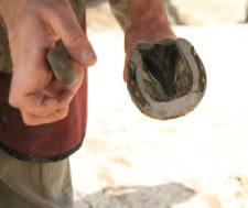
The team of 3D printing experts worked with horse podiatrists to scan Holly's feet and design the “horse-thotic” which aims to support the foot and encourage it to heal, whilst making Holly comfortable.
Holly has had laminitis for three years. Horse vet and farrier, Dr Luke Wells-Smith from the Equine Podiatry and Lameness Centre, said his team saw the 3D printed shoe CSIRO built for a race horse earlier this year and started to think about using 3D printing to rehabilitate lame horses.
"The new shoes will work to redistribute weight away from the painful areas of the laminitic foot and give Holly, and horses like her, the chance to recover," he said.
"Many attempts have been made in the past to cure laminitis but it’s the 3D scanning and design part of this process that is so exciting to us.
"Christmas is looking a lot merrier for Holly this year. She should be walking normally and without pain in just a few weeks," said Luke.
CSIRO's 3D printing expert, John Barnes, said scanning the hoof would allow them to manufacture a shoe that is the ‘perfect fit’ for these complicated foot diseases, giving the horse the best possible chance for rehabilitation," he said.
"We know that 3D printing has the potential to create so many advanced biomedical products, but rehabilitation of horses has been a completely new area of work for CSIRO.
"We’re glad that this technology is opening so many doors and is now helping to aid the rehab process for these animals and get them walking comfortably again," he said.
Tuesday, December 03, 2013
Limiting roughage access may limit fertility
Ensuring that mares have access to roughage throughout the day and night has beneficial effects on reproductive efficacy according to a recent report.
Horses have evolved as "trickle feeders" and are adapted to eating little and often. Modern management methods frequently interfere with this natural feeding routine, and have been implicated in causing welfare problems and health issues such as gastric ulcers and colic.
Now evidence has emerged to suggest that restricting the availability of ro ughageto just part of the day can be detrimental to breeding performance.
The study, “Temporal feeding pattern may influence reproduction efficiency, the example of breeding mares” by Haifa Benhajali and colleagues has been published in the journal PLOS One.
One hundred Arab breeding mares on the national breeding facility of Sidi Thabet, near Tunis, in Tunisia, were included in the study. They were randomly divided into two groups whose management differed only in the timing of availability of roughage. Mares were housed individually except for a period of six hours when they were turned out into a paddock. They had free access to water, a small amount of cut grass was given at midday, and 4kg of barley was fed overnight.
The “Continuous feeding pattern” group (CF) had access to hay both when turned out and when housed for the rest of the day and night. They received 5kg hay in the stable at night and 5kg hay when turned out. The “standard feeding pattern” group (SFP) received 10kg hay only when housed. So the total amount of roughage provided each day for both groups was the same.
The researchers found a significant difference between the two treatment groups. Those receiving hay throughout the day had fewer oestrus abnormalities and higher fertility. The conception rate in the CF mares was 81% compared with 55% in the SFP mares.
They conclude that “temporal patterns of feeding may be a major and underestimated factor in breeding.”
Temporal feeding pattern may influence reproduction efficiency, the example of breeding mares.
Benhajali H, Ezzaouia M, Lunel C, Charfi F, Hausberger M.
PLoS One. 2013 Sep 30;8(9):e73858.
Horses have evolved as "trickle feeders" and are adapted to eating little and often. Modern management methods frequently interfere with this natural feeding routine, and have been implicated in causing welfare problems and health issues such as gastric ulcers and colic.
Now evidence has emerged to suggest that restricting the availability of ro ughageto just part of the day can be detrimental to breeding performance.
The study, “Temporal feeding pattern may influence reproduction efficiency, the example of breeding mares” by Haifa Benhajali and colleagues has been published in the journal PLOS One.
One hundred Arab breeding mares on the national breeding facility of Sidi Thabet, near Tunis, in Tunisia, were included in the study. They were randomly divided into two groups whose management differed only in the timing of availability of roughage. Mares were housed individually except for a period of six hours when they were turned out into a paddock. They had free access to water, a small amount of cut grass was given at midday, and 4kg of barley was fed overnight.
The “Continuous feeding pattern” group (CF) had access to hay both when turned out and when housed for the rest of the day and night. They received 5kg hay in the stable at night and 5kg hay when turned out. The “standard feeding pattern” group (SFP) received 10kg hay only when housed. So the total amount of roughage provided each day for both groups was the same.
The researchers found a significant difference between the two treatment groups. Those receiving hay throughout the day had fewer oestrus abnormalities and higher fertility. The conception rate in the CF mares was 81% compared with 55% in the SFP mares.
They conclude that “temporal patterns of feeding may be a major and underestimated factor in breeding.”
For more details see:
Temporal feeding pattern may influence reproduction efficiency, the example of breeding mares.
Benhajali H, Ezzaouia M, Lunel C, Charfi F, Hausberger M.
PLoS One. 2013 Sep 30;8(9):e73858.
doi: 10.1371/journal.pone.0073858
Sunday, December 01, 2013
How risky is medicating joints?
Injecting medication directly into the joint is a common procedure for treating injuries in competition horses. It carries a risk of introducing infection into the joint. However, a recent report suggests that with adequate preparation serious consequences are uncommon.
Lewis Smith and colleagues examined clinical records of all horses given intra-articular injections by nine ambulatory veterinary surgeons over a five year period. The vets were based at a specialist equine practice in Newmarket England dealing mainly with Thoroughbred racehorses. Data relating to intra-articular injections for diagnostic purposes were not included in the study.
The site of injection was prepared using standard aseptic technique, but usually the hair was not clipped. All injections were made using scrupulous aseptic injection technique.
During the study period, 9456 injections were performed, including corticosteroids (92.3%) antibiotic (amikacin)( 94.8%) and polysulphated glycosaminoglycans (PSGAGs)(0.15%).
Twelve horses developed complications after medication. Four of those developed joint sepsis, but all returned to work after having the joint flushed.
Analysis of the data showed that administration of PSGAGs into the joint, was significantly associated with joint infection. Overall, intrasynovial medications that included amikacin were less likely to develop joint sepsis, but the difference was not significant if the PSGAGs were excluded from the analysis.
The authors concluded that the risk of sepsis being induced inadvertently following intrasynovial medication was extremely low. They advised that intrasynovial medication with PSGAGs should be avoided unless antimicrobials are given at the same time.
For more details see:
Risks of synovial sepsis following intrasynovial medication in ambuatory practice, 2006-2011: 9456 intrasynovial injections. Smith, L., Palmer, L., Shepherd, M., Steven, W.N., Dallas, R., Baldwin, G., Sommerville, G., Hawthorne, T. and Ramzan, P.
Equine Veterinary Journal (2013) 45, Suppl. 44, 6 (no11)
Friday, November 29, 2013
NEW LAMINITIS STUDY
A new
research project aiming to help horse owners reduce the impact of
laminitis is being undertaken by the Animal Health Trust (AHT) in
partnership with the Royal Veterinary College (RVC).
The study,
which will extend over four years, is being funded by World Horse
Welfare. It will take a closer look at management factors that may
contribute to the development or recurrence of laminitis within the
British horse and pony population. Through modifying these
contributing factors, it is hoped that horse owners can significantly
reduce the impact of this important welfare problem.
The AHT and
RVC plan to create a website where owners from all over the country
can register their horses and ponies and assist in the regular
gathering of information related to potential risk factors for
laminitis, over a period of two years.
This will
help establish a timeline of events and gain a better understanding of
the factors leading to laminitic episodes.
The study, to
be conducted by PhD student Danica (Dee) Pollard, will follow-up on
previous research conducted by Dr Claire Wylie in which factors such
as rapid weight gain, increasing time since last deworming, box rest
in the previous week and new access to grass in the past month
increased the risk of laminitis.
Dr Wylie’s
study also revealed that factors such as feeding of additional
supplements and transport in the previous week were associated with a
reduced risk of laminitis.
These are factors that could all be changed by the owner, and
this is why they are of particular interest to the new study.
Dee Pollard,
based at the AHT, said: “This will be a very exciting opportunity
for owners to be at the frontline of equine health research and
contribute to a study which aims to provide evidence-based
preventative strategies to combat laminitis.”
Horse owners
interested in taking part in the research project are asked to
register their interest via email to danica.pollard@aht.org.uk
Thursday, November 28, 2013
Acceptability of smaller microchips
Although microchips are widely used for identifying horses throughout Europe, there is still some resistance to their use, with questions being raised about stress during implantation, inflammation at the site of implantation, and reliability of detection.
In response to this criticism, microchips that are even smaller have been developed. They may be less stressful to implant, but do they work as well? Recent work has looked at whether such chips are reliable and if their implantation causes signs of stress.
The study, carried out at the Brandenburg State Stud at Neustadt (Dosse), Germany, was reported by Manuela Wulf and others in a recent edition of the Veterinary Record. Forty adult mares were implanted, on the left side of the neck, with a reduced-size microchip (10.9×1.6 mm). (Conventional microchips are 11.4×2.2 mm). Three different scanners were used to detect the microchips immediately, and on three further occasions up to 28 weeks after implantation.
The researchers found that scanners differed in their ability to read the microchips, although all scanners detected all chips on every occasion when scanned from the side of implantation. One scanner read all microchips successfully from both sides of the neck on four occasions up to 28 weeks after implantation. Two other scanners detected all of the chips from the side of implantation, but were less successful reading from the “wrong” side of the neck.
Did the horses find the implantation procedure stressful? The researchers monitored heart rate, heart rate variability and saliva cortisol levels during the implantation process in twelve of the mares. They also recorded the same information while pressing at the implantation site with a cannula without penetrating the skin. So each mare acted as its own control. They found a slight increase in heart parameters in both chip implantation and controls, but no change in cortisol levels.
The report's authors conclude that reduced-size microchips are highly reliable for identification of horses. “Compared with conventional microchips, the reduction in size did not impair readability. Microchip implantation is no pronounced stressor for horses.”
For more details see:
Reduced-size microchips for identification of horses: response to implantation and readability during a six-month period.
Wulf M, Aurich C, von Lewinski M, Möstl E, Aurich JE.
Vet Rec. 2013 Nov 9;173(18):451.
doi: 10.1136/vr.101824
Monday, November 25, 2013
Riding Arena Footing and Management Webcast
Ever wondered what's involved in providing a top quality arena surface, or wanted to know more about the materials to use?
Here's your opportunity. There's a free webcast on “Riding Arena Footing and Management” from MyHorseUniversity on November 26, 2013 at 7 PM ET.
Dr. Ann Swinker, Associate Professor in Equine Sciences, Penn State University and Horse Extension Specialist has been involved in the horse business for over 35 years. She will discuss the physical properties of the various footing materials that are available and the advantages and disadvantages. She will also consider the management of arena surfaces, the principles of maintenance and the signs that a surface needs to be changed or replaced.
You can register for the webcast now. Don't worry if you miss it; you can still catch it later, as it will be archived on the website and will continue to be available (free).
For more details go to ....
http://myhorseuniversity.com/resources/webcasts/ridingarenafooting
Benefit of targeted worming
Worming only those horses that need it can be cost effective, even taking into account the cost of performing faecal worm egg counts, according to research published in the Veterinary Record.
It is now widely acknowledged that a targeted approach to worm control is preferable to interval dosing regimes. Current recommendations are that only those horses carrying a moderate or high worm burden are treated; thus ensuring that worms are not exposed to anthelmintics needlessly.
Faecal worm egg counts (FECs) are used to determine which horses need (or don't need) treating. To many owners this may seem an unnecessary expense. However, recent work has shown that using FECs in this way helps reduce the overall cost of worming.
Hannah Lester, with colleagues at the Moredun Research Institute, and the Universities of Bristol, Liverpool and Edinburgh, monitored FECs at 3 monthly intervals over a nine month period. In all, 368 horses from 16 separate yards were involved in the study.
Horses with FECs greater than 200epg were treated, with pyrantel (in March and June) and ivermectin (in September). All horses received moxidectin/praziquantel in December.
The researchers compared the cost with that of a standard interval regime of two treatments with moxidectin and two of moxidectin and praziquantel - which is what had been common practice in the study population.
They estimated the cost of the two approaches by using average prices for anthelmintic products and faecal egg counts that they obtained off the internet. Even allowing for the cost of faecal egg count reduction tests (ie repeating the FEC after each treatment to check the anthelmintic had been effective) they found that, over the year, there was an average saving per yard of £294.44.
They conclude: “these findings support the notion that targeting anthelmintic treatments at those individuals with strongyle FEC of 200epg or greater facilitates a reduction in selection pressure for anthelmintic resistance. Moreover, the results show that such a strategy has a high chance of reducing the financial cost compared with that associated with more traditional interval treatment regimens, and horse owners should, therefore, be discouraged from the view that it is cheaper to treat all horses prophylactically over time.”
Read more: equinescienceupdate.com
Sunday, November 24, 2013
Emergency ventilation system developed
Respiratory or cardiovascular arrest in outdoor animals poses a huge challenge to veterinarians. Ventilation equipment is generally hard to operate and requires electricity and compressed air, and is not easily transportable.
Anaesthesiologists at the University of Veterinary Medicine, Vienna (Vetmeduni Vienna) have developed an inexpensive device that can be used to ventilate large animals. They report that it is easy to transport and can save animal lives in emergencies.
In work recently published in Equine Veterinary Education, the scientists confirm that their emergency ventilator is effective in horses.
Yves Moens is Head of the Vetmeduni’s Clinical Unit of Anaesthesiology and Perioperative Intensive Care Medicine. He and his colleagues have long been concerned by the number of horses that die avoidable deaths because of the lack of a suitable ventilation device. The device they designed is similar to the bellows used to inflate air mattresses but has been adapted by the addition of a manually operated expiratory valve.
Although it can only provide 2.5 litres of air, the researchers believed that it would provide sufficient ventilation if the bellows were activated several times in quick succession. They tested this idea on five anaesthetised Haflinger horses during castration surgery and showed that gradual ventilation with the 2.5 litre pump was sufficient to keep the animals alive.
“It improves the safety of large animals in the field, both during routine anaesthesia and in emergencies. It will also help veterinarians to provide emergency first aid in these circumstances and respect the guidelines for good practice", says Moens. “The respiratory pump is inexpensive and easy to use and will help veterinarians treat their patients in the field.”
Read more : equinescienceupdate.com
Saturday, November 23, 2013
How accurate is thermography of horses’ legs?

Recent work by Simone Westermann at the University of Veterinary Medicine, Vienna shows that the technique is surprisingly tolerant of variation in the position of the equipment, i.e. how far from the horse and at what angle to the animal the infrared camera is held.
In fact, the results were almost completely unaffected by 20° changes in camera angle and increases of up to 50 cm in the distance of the camera from the animal. At a distance of 1m from the horse a 20° change in camera angle corresponds to about 35 cm. This represents the effective horizontal tolerance in positioning of the camera. As Westermann says, “vets should have little difficulty in remaining within this limit, so the method is applicable in practice”.
Surprisingly, the results showed that horses’ left and right forelimbs show minor differences in temperature and Westermann cautions that “it might be important to take these into account before reaching a final diagnosis.”
The technique is thus reliable and robust, at least in terms of variation in where the camera is located.
However, it turned out to be extremely sensitive to even very gentle drafts. A wind speed of less than 1 m/s causes a drop in measured temperature of about 0.6°C, while winds of 1.3-2.6 m/s cause a drop of 1.5°C and winds of 3-4 m/s cause a drop of 2.1°C. The discrepancies are more than sufficient to lead to a wrong diagnosis, although even the highest wind speed tested is hardly perceptible: it would barely cause leaves on trees to move.
Westermann is keen to note the relevance of her work for vets who work on horses. As she says, “It turns out that it is not too important to be sure that the camera is in exactly the correct position before taking measurements. But it is essential to perform thermography on horses in a room that is completely free of draughts. If you don’t, your diagnosis will be completely unreliable.”
More information : equinescienceupdate.com
Thursday, November 21, 2013
Antibacterial action of honey
Wounds to the lower limbs of horses can prove challenging to manage. Recently there has been a growing interest in the use of honey in such cases.
Not all honey is the same. Its antibacterial quality depends on the type of honey and the conditions under which it was harvested and processed. Most honey contains hydrogen peroxide, which has antibacterial properties. Some types of honey contain other active components. For example, manuka honey is believed to have antibacterial properties due to high concentrations of methylglyoxal, a compound usually found in only low quantities in other types of honey.
Manuka honey, produced by bees foraging on manuka plants (Leptospermum scoparium), native to Australia and New Zealand, has been the subject of considerable research. Honey from other sources is often used in practice, but there has been little research into how effective it is.
Researchers at the University of Glasgow have been examining various different types of shop-bought honey to determine if they were free from bacterial contamination and suitable for use on equine wounds. They also investigated the effect of various examples of uncontaminated honey on the growth of equine pathogens, and found that, in laboratory tests, certain varieties of honey are able to inhibit bacterial growth even at very low concentrations.
They found that many commercial sources of honey were contaminated with bacteria. The most commonly identified contaminating organism was Bacillus spp. However, potentially pathogenic organisms (Proteus and Enterobacteraceae) were identifed in two honey samples.
Uncontaminated honey samples were subjected to further investigation to assess their antibacterial properties. Eight of the eleven samples tested were effective against all 10 bacterial isolates at concentrations from 4 to 16%. Overall, medical grade Manuka honey and a locally produced heather honey performed best.
The researchers conclude that many honeys have antimicrobial properties, and may be effective in the treatment of wound infections. They note that the concentrations at which honey samples inhibited microbial growth were much lower than is likely to occur at the surface of an infected wound treated with honey.
However, they advise that “the use of shop-bought honey on wounds should be avoided, as contamination with potentially pathogenic microbes appears to be common. Honey sourced within the UK is as, and in some cases more, effective than medical grade honey sourced in New Zealand.”
Read more at equinescienceupdate.com/articles/aah.html
Wednesday, November 20, 2013
New laminitis research findings
Laminitis continues to be a significant welfare problem of horses and ponies, causing widespread suffering. Investigations into the underlying causes and disease processes involved in the condition are ongoing.
The benefits of feeding a balanced diet alongside appropriate forage, grazing restriction and regular low intensity exercise whenever clinically possible in the management of laminitis prone horses and ponies have been highlighted by new research.
Four separate studies have shed new light on the possible role of grass fructan in the development of laminitis; the influence of water temperature when soaking hay to reduce the water-soluble carbohydrate content; a possible link between recurrent laminitis and reduced anti-inflammatory capacity, and the potential anti-inflammatory benefits of exercise.
Read more at equinescienceupdate.com
Tuesday, November 19, 2013
Controlling forage intake with hay nets
Horses have evolved to spend much of the day grazing. However, modern systems of horse management often restrict the time available. This may contribute to problems such as gastric ulceration and can result in behavioural problems.
Sometimes it is desirable to reduce the intake of roughage – either to make the horse's ration last longer, or to limit its overall intake.
Recent research from the University of Minnesota shows that using hay nets with smaller holes is effective for limiting the rate of roughage ingestion in horses.
Eight horse were involved in the study. They were were housed in individual box stalls, and fed hay off the floor (control treatment) or from hay nets with one of three different sized holes. The mesh size ranged from 15.2cm (large), to 4.4cm (medium) and 3.2cm (small).
During the trial period, hay was available for two four-hour periods each day.
Horses were allowed to become accustomed to each type of net for 2 days, before intake was recorded over three days. They then had two days of a wash out period during which they were as a group in an outdoor paddock.
The researchers found a significant difference in the rate of consumption between all treatment groups. Horses fed hay off the floor (control) consumed hay at the rate of 1.49kg/hr. Consumption of hay from hay nets was 1.33kg/hr, 1.11kg/hr, 0.88kg/hr for large, medium and small sized holes respectively.
They found no difference between the large mesh net and control for the amount of hay consumed (both 95%% of hay offered); but medium and small hay nets restricted hay intake to 89% and 72% respectively.
They conclude that their results demonstrate that the medium and small sized nets were effective in decreasing both rate and amount of forage consumed by adult horses.
More information at equinescienceupdate.com
Subscribe to:
Posts (Atom)