Monday, December 20, 2021

Fragile Foal Syndrome not confined to Warmbloods

 Fragile Foal Syndrome (FFS) is a lethal genetic disease of connective tissue which has been reported most frequently
in Warmbloods. However, a recent study has found that the genetic defect responsible is present across a range of other breeds.
 

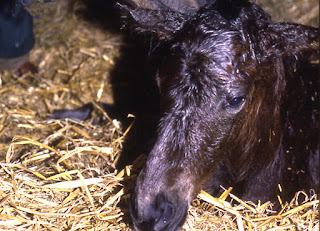
Affected foals are typically aborted during late gestation or born as non-viable foals. If alive at birth, they tend to have problems such as fragile skin, skin defects, hyperextension of the joints and difficulty breathing, and generally require euthanasia within days.


FFS has been shown to be an autosomal recessive genetic condition. Carrier animals with one copy of the defective gene (PLOD 1 c.2032 G>A) will be normal, but if mated with another carrier may produce an affected foal.


It is now known that the condition is not confined to Warmblood horses.  


Research by Katie Martin and colleagues at Etalon diagnostics (a company that offers genetic testing), together with Dr Samantha Brooks at the University of Florida Gainesville and Dr Scott Mclure, of Midwest Equine, Iowa, found that the genetic defect occurs across other horse populations.


The team examined samples from 7343 horses from various breeds or type of horse.

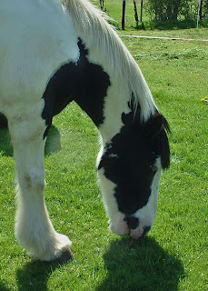
The defective gene occurred in 5.32% of Warmblood type horses.  In other affected breeds it was less than 1%. They found no sign of the defect in Arabians, Iberian or Thoroughbreds.


Studies of the frequency of the defect in aborted or stillborn foals are lacking, so

the potential economic effect of FFS on the horse breeding industry is not known.


The researchers suggest that pre-breeding testing should be used to inform the breeding program – avoiding breeding two carriers, together to reduce the frequency of the FFS gene in the population, and reduce the number of lost pregnancies.


For more details, see:


Fragile Foal Syndrome (PLOD1 c.2032G>A) occurs across diverse horse populations

Katie Martin, Samantha Brooks, Micaela Vierra, W. Tyler Lafayette, Scott McClure, Meredith Carpenter and Christa Lafayette

Animal Genetics (2021) vol 52, p137.

https://doi.org/10.1111/age.13020

Sunday, December 19, 2021

Reconstructing prehistory from a teaspoonful of soil

Yukon Photo credit Tyler Murchie
Prehistoric horses survived longer in North America than previously thought, according to a new study.

Researchers at McMaster University, Hamilton, Canada, used DNA capture-enrichment techniques, that they had developed previously, to find ancient DNA from plants and animals in as little as a teaspoonful of soil. The soil for the study was taken from core samples collected from the permafrost at four sites in the Klondike region of central Yukon, in northwest Canada. 


The work is published in the journal Nature Communications.


Tyler J. Murchie  and co-authors explain that environmental samples, such as soil, contain fragments of genetic material. Most ancient environmental DNA (eDNA) is broken down by bacteria or by physical or chemical processes. However, some of this eDNA becomes bound to sedimentary minerals, which protects it, especially when it is frozen. 


By extracting and analysing this sedimentary ancient DNA (sedaDNA), the research team was able to build up a picture of the plant and animal ecosystems from 30,000 to 4000 years ago. 


By analysing the DNA, the research team could rebuild the fluctuating animal and plant communities at different time points. Of particular interest was the Pleistocene-Holocene transition, an unstable climatic period 11,000-14,000 years ago when a number of large species such as mammoths, mastodons and sabre-toothed cats disappeared.

The analysis reveals that mammoths and horses were already in steep decline prior to the climatic instability, but they did not immediately disappear due to human overhunting as previously thought. In fact, the DNA evidence shows that both the woolly mammoth and North American horse persisted until as recently as 5,000 years ago.

The authors explain that through the early Holocene (starting about 11,000 years ago), the Yukon environment continued to experience massive change. Formerly rich grasslands - the “Mammoth Steppe”- were overrun with shrubs and mosses, species no longer held in check by large grazing herds of mammoths, horses and bison. Today, grasslands do not prosper in northern North America, in part because there are no megafaunal “ecological engineers” to manage them. 

“The rich data provides a unique window into the population dynamics of megafauna and nuances the discussion around their extinction through more subtle reconstructions of past ecosystems” says evolutionary geneticist Hendrik Poinar, a lead author on the paper and director of the McMaster Ancient DNA Centre.

This work builds on previous research by McMaster scientists who had determined woolly mammoths and the North American horse were likely present in the Yukon approximately 9,700 years ago. Better techniques and further investigation have since refined the earlier analysis and pushed forward the date even closer to contemporary time. 

“Now that we have these technologies, we realize how much life-history information is stored in permafrost,” explains Tyler Murchie, a postdoctoral researcher in McMaster’s Department of Anthropology and a lead author of the study.

“The amount of genetic data in permafrost is quite enormous and really allows for a scale of ecosystem and evolutionary reconstruction that is unparalleled with other methods to date” he says.

“Although mammoths are gone forever, horses are not” says Ross MacPhee of the American Museum of Natural History, another co-author. “The horse that lived in the Yukon 5,000 years ago is directly related to the horse species we have today, Equus caballus. Biologically, this makes the horse a native North American mammal, and it should be treated as such.” 

Scientists also stress the need to gather and archive more permafrost samples, which are at risk of being lost forever as the Arctic warms.


For more details, see:


Collapse of the mammoth-steppe in central Yukon as revealed by ancient environmental DNA

Tyler J. Murchie, Alistair J. Monteath, Matthew E. Mahony, George S. Long, Scott Cocker, Tara Sadoway, Emil Karpinski, Grant Zazula, Ross D. E. MacPhee, Duane Froese & Hendrik N. Poinar 

Nature Communications (2021) vol 12, Article number: 7120 


https://doi.org/10.1038/s41467-021-27439-6


For a video presentation, see:

https://player.vimeo.com/video/654303546

Saturday, December 18, 2021

A cheaper alternative for URT scoping

Is there a cheaper alternative to the traditional endoscopes used for examination of the upper respiratory tract (URT) of horses?

Flexible endoscopes have proved invaluable for inspecting the inner workings of the equine respiratory system for over 40 years. The equipment is expensive and so is not always readily available. 


Borescopes are commonly used in industry for tasks such as the inspecting the inside of engines and other confined spaces.  A wide variety of types are available, most considerably less expensive than medical grade endoscopes. 


Researchers in the Faculty of Veterinary Medicine, Ghent University, investigated the suitability of a flexible and steerable borescope connected to a smartphone for examining upper respiratory tract problems in horses. Zoe Neuchermans and her colleagues in the department of Large Animal Internal Medicine conducted a study involving a series of clinical cases. Horses were inspected first with a flexible borescope and then with the standard flexible endoscope.


They made digital recordings of upper respiratory tract including structures of interest (depending on the clinical case). These were later reviewed by an observer who did not know which device had been used to make the recording. The work was presented at the British Equine Veterinary Association Congress 2021 and has been published in the Equine Veterinary Journal.


The authors report that “borescope and endoscope grading scores for pharyngeal lymphoid hyperplasia, recurrent laryngeal neuropathy and tracheal mucus were identical in 88/100, 93/100 and 48/59 horses, respectively. The remainder differed only one (sub)grade.”


The only problem they reported was that they had to replace the borescope after about 45 examinations as the steering mechanism started to fail ,which limited the field of view.


Although their study was limited to only one of the many types of borescope available, the researchers came to the conclusion that a flexible, steerable borescope, connected to a smartphone, provides a cheap alternative to perform URT endoscopy in horses.


For more details, see: 


Diagnosis of upper respiratory tract disorders in horses using a cheap, flexible and steerable borescope

Z. Neuckermans; E. Paulussen; L.-M. Verhaeghe and G. van Loon.

Equine Vet J (2001) Vol 53, S55, p22-23

 https://doi.org/10.1111/evj.33_13492

Friday, December 17, 2021

Fungal changes in gut of horses with grass sickness

Horses with Grass Sickness have differences in the population of fungi (“mycobiota”) present within in the gut
compared with healthy horses, a recent study has revealed.

Equine grass sickness (EGS, also known as equine dysautonomia) damages the autonomic nervous system – the part of the horse’s nervous system that controls involuntary functions. Its effects are particularly apparent in the gastro-intestinal tract. Clinical signs range from depression, with difficulty eating and swallowing, to reflux of stomach contents, weight loss and death. 

The exact cause is unknown, but researchers now know a lot about risk factors to be avoided and believe that a type of toxin is involved.  Is EGS is associated with ingestion of mycotoxin-producing fungi? 

Researchers, led by Bruce McGorum at the University of Edinburgh, conducted a study to identify potential causal fungi. They analysed the mycobiota within the gut contents of horses with grass sickness and compared their findings with samples taken from horses that died from reasons other than gastro-intestinal or neurological problems. They also analysed faecal samples from unaffected horses grazing the same pasture as horses with grass sickness.

They found a very rich and diverse gastro-intestinal mycobiota in all the horses. The mycobiota appeared to be richer (ie containing more different types of fungi) and more diverse (a more even distribution of the types of organism) in grazing horses. Mycobiota richness was greater in horses with EGS. 


The research team identified 56 key “phylotypes” that were more common in EGS samples. Many of these key phylotypes were extremophiles (organisms able to survive in extreme environments) or were considered likely to produce extrolites (secreted metabolites) that might have toxic effects.

These changes in the mycobiota have not been shown to cause EGS. Further work is needed to determine whether neurotoxic extrolites from these key phylotypes play a role in causing grass sickness.


For more details, see:

Equine grass sickness (a multiple systems neuropathy) is associated with alterations in the gastrointestinal mycobiome

Bruce C. McGorum, Zihao Chen, Laura Glendinning, Hyun S. Gweon, Luanne Hunt, Alasdair Ivens, John A. Keen, R. Scott Pirie, Joanne Taylor, Toby Wilkinson & Gerry McLachlan 

Animal Microbiome (2021) vol 3, Article number: 70 

https://doi.org/10.1186/s42523-021-00131-2

Monday, December 13, 2021

Search for a test to predict horses at risk of catastrophic injuries

In the ongoing quest to make racing safer for horses, researchers have been looking for a blood test to identify horses at risk of catastrophic fractures.

Other than sudden high energy trauma resulting from falls in jump racing, most racing fractures are thought to be fatigue injuries which result from cumulative stresses and strains.


Advanced imaging techniques, such as computed tomography (CT), magnetic resonance imaging (MRI), and positron emission tomography (PET) scans, have been used to identify damage that might lead to catastrophic fractures. However, such methods would be too expensive and impractical for routine use.


Most catastrophic fractures are found to have underlying pre-existing damage. The associated inflammation results in changes in the activity of various components of the inflammatory process, So checking for inflammatory markers might give warning of potential risk.


Dr Allen Page and colleagues at the University of Kentucky’s Maxwell H Gluck Equine Research Center, hypothesised that analysis of messenger RNA expression would detect significant changes in horses at risk for a catastrophic injury.


Their study involved blood samples collected from Thoroughbred horses in five racing jurisdictions across the United States. The work is published in the Equine Veterinary Journal.


They looked at 21 genes potentially associated with bone inflammation and remodelling,  and compared the levels of gene expression in horses that had suffered a catastrophic fracture with those in horses that had raced without injury.


Three markers, - insulin like growth factor 1 (IGF-1), matrix metalloproteinase 2 (MMP 2) and IL-1 receptor antagonist (IL1RN) - showed a marked difference between injured and non-injured horses. The researchers found that together, the three markers correctly identified horses at risk of catastrophic injury 76% of the time (and correctly excluded horses from being at risk 88% of the time)


They conclude that “Analysis of mRNA expression of specific genes in the future may be considered as an economical, accessible and non-invasive means by which horses at risk for catastrophic injury can be identified.”

For mor details, see:

Expression of select mRNA in Thoroughbreds with catastrophic racing injuries.
Page AE, Adam E, Arthur R, Barker V, Franklin F, Friedman R, Grande T, Hardy M, Howard B, Partridge E, Rutledge M, Scollay M, Stewart JC, Vale A, Horohov DW.
Equine Vet J. 2022; vol 54(1) pp63-73. 

https://doi.org/10.1111/evj.13423

Friday, November 26, 2021

Evaluating chronic pain in horses - podcast

In a podcast recorded recently, Dr. Kelly Diehl, Senior Director of Science and Communication at the Morris Animal Foundation spoke to Drs. Janny de Grauw and Diane Howard about the challenges associated with the detection and measurement of chronic pain in horses. 

Osteoarthritis, a leading cause of chronic pain in horses, is often not recognized by owners and so goes undertreated. 


Dr. de Grauw and Dr. Howard also discussed how they developed their pain assessment tool and how they hope this will help thousands of horses suffering from chronic pain.

Under Dr. de Grauw’s supervision, Howard developed a 15-item questionnaire based on changes in horse behaviour through interviews with owners of horses diagnosed with osteoarthritis. The questions cover posture, facial expressions, movement and behaviour.

“As veterinarians, we want to treat horses with painful and debilitating conditions like OA as effectively as possible,” said de Grauw. “How well we can manage their condition critically relies on recognition of subtle signs of (worsening) pain by owners and caregivers, who can then seek help.”

Listen to the podcast here:

https://podcasts.apple.com/us/podcast/evaluating-chronic-pain-in-horses/id1440087311?i=1000541242539

Thursday, November 25, 2021

Alternative approach to colic ops


Standing flank laparotomy may be a suitable option for surgical treatment of some cases of equine colic, a recent study has concluded.

The standard surgical approach for colic is through an incision in the ventral midline, with the horse under general anaesthesia. General anaesthesia poses additional risks over and above those of the surgery itself, and the personnel and equipment required increases the cost.


Operating through a flank incision would avoid the need for general anaesthesia and should be less expensive.


Marco Lopes and co-workers conducted a retrospective analysis of clinical records of 37 equids (horses, ponies and a donkey) treated using a standing flank laparotomy at five hospitals, between 2003 and 2020. Their findings are published in the Equine Veterinary Journal.


They did not include cases of small colon impaction in their analysis, as previous work had already shown that such cases could be dealt with using a flank incision.


Financial considerations were the main reason for the choice of technique. However, in ten cases of nephrosplenic entrapment the surgeon preferred a flank incision.


In seven animals the decision for euthanasia was taken immediately as they were found to have an untreatable condition, or the likelihood of survival was very poor.


Twenty of the 30 horses found to have a treatable condition survived. These conditions included small intestine inflammation or impaction, large colon displacement (especially nephrosplenic ligament entrapment) and sand impaction, 


The authors identify limitations of the technique:


  • the horse must stand still during the operation, so standing flank laparotomy cannot be performed in horses with severe colic that cannot be adequately controlled medically
  • additional peritoneal analgesia may be needed
  • access to the peritoneal cavity and the abdominal organs is not as good as with a ventral midline incision.
  • a second incision on the opposite flank may be needed
  • surgeons are not as familiar with standing flank laparotomy 
  •  scars in the flank are more likely to be seen than those in the ventral midline.


They conclude that this series of cases indicates that standing flank laparotomy is a viable approach for abdominopelvic exploration in some horses with colic attributed to conditions of the small intestine, caecum, large colon and peritoneum.


For more details, see: 


Standing flank laparotomy for colic: 37 cases

Marco A. F. Lopes, Joanne Hardy, Kelly Farnsworth, Raphael Labens, W. Y. Eunice Lam, Erik Noschka, Tiago Afonso, Claudia Cruz Villagrán, Luiz C. P. Santos, Montague Saulez, Gal Kelmer

Equine Vet J (2021)


https://doi.org/10.1111/evj.13511


Wednesday, November 24, 2021

Investigating a possible treatment for melanoma

Initial indications are that betulinic acid may prove to be a useful topical treatment for early-stage melanomas in horses. 

Melanomas are tumours of the pigment-producing melanocytes, and are among the most common equine tumours, occurring particularly in grey horses. Typically, they first appear as small, pigmented nodules, often under the tail. They usually grow slowly for years, but may infiltrate surrounding tissues, or appear elsewhere in the body.


Betulinic acid (BA) is a natural compound, found in the bark of several species of plants, such as the

white birch (Betula pubescens) after which it was named. It has been shown to have several potentially-useful pharmacological effects. Previous work has shown that BA can induce death (apoptosis) of melanocytes in cell culture. 


Research conducted in Austria looked at whether topical treatment with betulinic acid, or NVX-207, (a betulinic acid derivative) could be used to treat early cases of melanoma.


Lisa A. Weber and colleagues conducted a small, pilot study involving eighteen Lipizzaner mares with cutaneous melanomas (all having a diameter of no more than 15mm).


The horses, on a stud farm in Austria, were divided into three treatment groups: placebo cream; 1% BA cream; and 1% NVX-207 cream.


The research team treated a maximum of two tumours per horse. After topical application of the cream, the melanomas were covered to prevent it being rubbed off. The study extended over 13 weeks.


The researchers assessed progress with clinical examinations, measuring the tumours, monitoring them and the surrounding clinically normal skin, assessing the horses’ behaviour during cream application, and monitoring haematologic and blood biochemistry profiles. 


All horses tolerated the topical drug application well and did not object to it being applied. Two horses had mild colic during the study, which resolved with medical treatment. Both horses had a previous history of colic and the authors consider it very unlikely that the colic was related to the topical melanoma treatment.


The topical therapy resulted in part in clinically visible and measurable changes in small melanocytic lesions, which were reflected in skin depigmentation and reduction in tumour diameters and volumes. 


The researchers found a beneficial effect after treatment with BA towards the end of the treatment period. A few tumours in the placebo group also showed a decrease in size.


They conclude: “The results presented in this pilot study indicate that topical treatment of early-stage equine melanoma with 1% BA and 1% NVX-207 twice a day over a period of 13 weeks is feasible and safe.” 


“Especially after BA application, positive effects were observed toward the end of the treatment interval. This suggests that this approach might be a potential therapy for early-stage equine melanoma and, thus, reduce the health risks associated with the possible malignant degeneration of the tumours.”


However, the authors are unable to recommend the current treatment protocol for general use as the long treatment duration could lead to poor owner compliance. They suggest that modification of the pharmaceutical formulations may improve the clinical outcome and reduce treatment periods.


For more details, see:


Effects of Topically Applied Betulinic Acid and NVX-207 on Melanocytic Tumors in 18 Horses.

Weber, L.A.; Delarocque, J.; Feige, K.; Kietzmann, M.; Kalbitz, J.; Meißner, J.; Paschke, R.; Cavalleri, J.-M.V. 

Animals 2021, 11, 3250. 

https://doi.org/10.3390/ani11113250

Tuesday, November 23, 2021

Looking for adverse effects of pergolide


Pergolide is widely used in the treatment of PPID (Pituitary Pars Intermedia Dysfunction, or equine Cushing’s disease). The drug is also used in humans, where it has been associated with side effects, involving fibrotic lesions of the atrio-ventricular valves in the heart.

Heidrun Gehlen and colleagues in the Department of Veterinary Medicine, at the Free University of Berlin, conducted a study to see if pergolide causes similar problems in horses. 
 
Twenty-three horses of various breeds, aged between 19 and 30 years old, were included. All had been diagnosed previously with PPID, based on ACTH (adrenocorticotrophin hormone) concentration.
 
Twelve horses had been receiving pergolide for between 14 days and 6 years before the start of the study. These comprised the treatment group and continued to receive pergolide. Eleven other horses received no pergolide.
 
The researchers performed a complete echocardiographic exam on all horses, which showed that they were free of cardiovascular diseases, including the absence of valvular defects. 
 
Follow up examinations were performed on nine horses in the treatment group and five of the non-treatment group between 3 and 8 months later.
 
The researchers compared the findings in PPID horses being treated with pergolide with the untreated PPID horses.
 
They found that treatment with pergolide did not affect the ventricular function nor induce valvular disease. Measurements taken in the follow-up exam did not differ from those taken initially in both groups. None of the 12 pergolide-treated horses developed valvular regurgitation. 
 
The work is reported in the Journal of Veterinary Science. The authors conclude: “The main result of our small orientation study was that treatment with pergolide of a duration between 3 and 8 months in a horse population with confirmed PPID did not seem to affect the left ventricular function assessed by TDE and STE [tissue Doppler and two-dimensional speckle tracking echocardiography]. Furthermore, none of the horses developed valvular regurgitation in the observation period.”
 
They add that further studies are needed, with a larger population and longer follow-up period. 
 
For more details, see:
 
Preliminary study on the effects of pergolide on left ventricular function in the horses with pituitary pars intermedia dysfunction
Heidrun Gehlen,  Judith Fisch,  Roswitha Merle and Dagmar S. Trachsel
J Vet Sci. 2021 Sep;22(5):e64. 
https://doi.org/10.4142/jvs.2021.22.e64 

Monday, November 15, 2021

New high-performance riding simulator

A novel horse-riding simulator offers new possibilities for rider training and welfare of the ridden horse.
The simulator was developed at the Department of Mechanical Engineering at Lappeenranta-Lahti University of Technology (LUT) in Finland, as part of a project to monitor body and brain behaviour of both professional and non-professional riders.


The new equipment will be unveiled at the Saddle Research Trust’s 4th International Conference, to be held online on Saturday 11th December 2021. Professor Heikki Handroos PhD, from the Lappeenranta-Lahti University of Technology will show how engineering science has been applied to develop the next generation horseback riding simulator.


“This realistic horseback riding simulator can benefit riders with different skills in many ways,” said Professor Handroos.


“Beginners can learn how to sit on the saddle during the basic gaits before starting to ride a real horse, which reduces injury risk and improves the horse welfare. The simulator can also carry heavier riders to help them to access the hobby with reduced welfare risks. For more advanced riders the technology will enable them to practice and enhance their skills as often as they wish.”


The high-performance novel robotic motion platform has been designed to provide the necessary motion capabilities for the simulator in all gaits, including jumping. The current test simulator is programmed with motions which were measured from advanced level dressage and show jumping horses whilst being ridden by advanced riders. 


The new simulator also has promise as a hippotherapy tool. “It has the potential to enable the ideal gait pattern to be programmed for each patient,” explained Professor Handroos. “We should also be able to use sensors to monitor the rider, while the simulator is performing different gait patterns. The same sensor technology could also be used in riding schools to monitor the learning curves of riding students.”


“Our next project is going to be on sensing the rider’s bio-signals when riding the simulator and intelligent processing of sensor data to assess the progress of riding school students or hippotherapy patients.”


To find out more about the Saddle Research Trust Conference go to:

https://www.saddleresearchtrust.com/conferences/


For more details, see:


Monitoring of the human body and brain behavior using optical motion capture system and EEG utilizing horseback riding simulator: an extended case study

Alina Byzova, Hamid Roozbahani, Heikki Handroos, Nils Hakansson, and Hamid M. Lankarani

J Phys Ther Sci. 2020 Jan; 32(1): 85–91. 

https://doi.org/10.1589/jpts.32.85


Thursday, October 21, 2021

Insights into strangles in the UK

 A new study from the Royal Veterinary College (RVC), analysing laboratory diagnoses of strangles across the UK, paves the way for an improved understanding of the spread and control of strangles to reduce the impact of this devastating disease.

Strangles is a contagious upper respiratory tract infection, caused by the bacterium Streptococcus equi, which can affect horses, ponies and donkeys of any age, breed or sex with younger horses typically more severely affected. It is also one of the most prevalent infectious diseases amongst horses and ponies worldwide, carrying a very high welfare burden with up to 100% of horses in outbreaks becoming affected.


This study, funded by The Horse Trust, brought together an international team from the RVC, the University of Melbourne, jDATA, Intervacc AB, Wellcome Trust Sanger Institute and the British Horseracing Authority. A full report of the work is published in the Veterinary Record.


Researchers analysed data from seven UK diagnostic laboratories between January 2015 and December 2019, finding that 1,617 laboratory diagnoses of strangles were recorded during that time. However, considering the number of potentially undiagnosed horses, the true number of equids affected by strangles is thought to be much higher.


Importantly, the findings of this study begin to quantify the occurrence of strangles within the UK and guide veterinary surgeons in their approach to disease diagnosis. This includes not ruling out a strangles diagnosis when a horse or pony presents with more general clinical signs of nasal discharge, with or without fever, in the absence of abscessation or swelling of the submandibular and retropharyngeal lymph nodes. More generally, the study suggested that the description of ‘classical’ and ‘atypical’ clinical signs should be revised.


The study also provides a valuable resource for UK horse owners in the form of an online tool to identify if strangles outbreaks have occurred in their area, or a region they may be travelling to with their horses. This resource is actively updated – so if a region is currently experiencing higher numbers of strangles diagnoses, owners can stay informed and subsequently heighten their biosecurity and hygiene protocols. This will help to reduce the spread of strangles and ultimately the impact it can have on yards, owners and horses.


This resource is actively updated meaning that if a region is currently experiencing higher numbers of strangles diagnoses, owners can stay informed and subsequently heighten their biosecurity and hygiene protocols. This will help to reduce the spread of strangles and ultimately the impact it can have on yards, owners and horses.


The publication also reinforces the benefit of a united front for strangles research and how through laboratories, veterinary practices and owners working together, we can provide much more detailed insights into the disease, leading towards safeguarding the health of our horses.

Abigail McGlennon, PhD student in the Department of Pathobiology and Population Sciences, Royal Veterinary College, and lead author of the report, said: “Prior to the development of the Surveillance of Equine Strangles network in 2018, there was limited information available about strangles diagnoses in the UK. This publication highlights the prevalence of strangles in the UK and the variation in signs that infected horses show. The results of this five-year surveillance study enable the continued development of evidence-based recommendations within the equine industry to help reduce the spread of strangles and keep our horses healthy and happy.” 


For more details, see the full (open access) article:


Surveillance of strangles in UK horses between 2015 and 2019 based on laboratory detection of Streptococcus equi. 

McGlennon A, Waller A, Verheyen K, Slater J, Grewar J, Aanensen D, Newton R. 

Vet Rec. 2021;e948.  

 https://doi.org/10.1002/vetr.948


To visit the Surveillance of Strangles website, go to :

https://app.jshiny.com/jdata/ses/sesview/

Dangers of equestrian activities severely under-appreciated say US researchers.

  A recent study has found that the risk of an injury, requiring hospital admission, is higher for horse riding than for other potentially risky sporting activities, such as football, motor racing, or skiing.

Kevin Mutore and colleagues examined data supplied from level I and II trauma centers to the US National Trauma Data Bank (NTDB), on injuries sustained by adults while horse riding between 2007 and 2016. 


They retrieved details of 45,671 patients with equestrian injuries for this period. Data were incomplete for 20,880 patients, leaving 24,791 for inclusion in the analysis. The average age of those injured was 47, with almost equal proportions of men and women. 


Analysis showed that the most common site of injury was the chest, (37%) followed by arms and legs (26.5%). Head and neck injuries, although occurring less commonly (23%), were the most likely to prove fatal.


Severe neurological damage, classified as a Glasgow Coma Scale (GCS) score of 3–8, was observed in 888 (3.5%) patients. The GCS is a clinical scale used to measure a person's level of consciousness after a brain injury. It ranges from 3 (deep coma) to 15 (normal consciousness.) 


Riders with head and neck injuries were 44 times as likely to die as those with arm/leg injuries, while those with chest and abdominal injuries were around 6 times as likely to do so. 


The researchers point out that the study only included data from patients treated at US trauma centers that reported data to the NTDB. Nevertheless, the findings prompt them to conclude that: “Equestrian-related injuries are a frequently ignored public health issue.”


They go on to say: “When taken together, these data suggest that the dangers of equestrian activities have been severely underappreciated. When controlled for hours of activity, horseback riding resulted in a higher proportion of hospital admission than other higher risk activities like skiing.”


Protective gear can save lives, but is not always worn, they highlight. “Studies have shown that a large fraction of riders involved in equestrian injuries were not wearing helmets at the time of their accident. It stands to reason that raising awareness of the possible injuries and increasing preventive measures to protect against head injuries would significantly reduce mortality.”


They conclude: “We suggest that preventive measures and campaigns should be instituted to highlight safety practices. Implementing the consistent use of personal protective equipment, such as helmets and vests, will provide added protection to all riders (working or leisure) while on horseback. It is also imperative that medical professionals examine patients injured during horseback riding for head and neck injuries as these contribute to the highest mortality.”


Full details of the research are available in the open access paper published in the online journal Trauma Surgery & Acute Care Open.

See:


Mutore K, Lim J, Fofana D, Torres-Reveron A, Skubic JJ

Hearing hoofbeats? Think head and neck trauma: a 10-year NTDB analysis of equestrian-related trauma in the USA

Trauma Surgery & Acute Care Open 2021;vol 6: e000728. 

http://dx.doi.org/10.1136/tsaco-2021-000728

Tuesday, October 19, 2021

Free Equitation Science Conference

There’s just time to register for the International Society for Equitation Science conference, which will be held online on 20th – 22nd October. Thanks to the generosity of the sponsors, it is being offered completely free of charge.


With the theme of Advancing Equestrian Practice to improve Equine Quality of Life, the conference is open to all with an interest in improving the welfare of the horse in its interactions with us. 


It aims to bring together horse riders, pony clubs and riding clubs, coaches and trainers, and equine students as well as academics, researchers and scientists to show how using evidence-informed approaches can advance equestrian practice and improve equine quality of life. 


For more details, see:


https://equitationscience.com/conferences/

Thursday, October 14, 2021

Hendra virus found in flying foxes across Australia


Scientists at CSIRO, Australia’s national science agency, have identified a new type of Hendra virus inflying foxes. The findings have been published in Virology Journal.

Hendra virus (HeV) causes lethal disease outbreaks in horses and humans. Flying foxes (fruit bats) provide a wildlife reservoir for the virus. Hendra virus can be transmitted from flying foxes to horses, and from horses to people. 


Previous studies have found the virus in flying foxes in Queensland and parts of New South Wales. They suggested that the black and the spectacled flying foxes were the primary carriers of Hendra virus. 


After monitoring flying fox samples from 2013-2021, researchers at CSIRO’s Australian Centre for Disease Preparedness (ACDP) found the new genetic type (designated HeV-g2)  in grey-headed flying foxes in Victoria and South Australia, and in the little red flying fox in Western Australia. 


The new genetic type was first detected in a flying fox sample from 2013, but with technology available at the time the researchers could not fully analyse its genome sequence to confirm its identity and understand its significance.


By piecing together the new virus’ genome from several flying fox samples since then using the latest technology, they discovered it was indeed a new type of Hendra virus. Ninety-eight flying foxes tested negative to the original Hendra virus, but 11 were found to carry genetic material indicative of HeV-g2.


The findings show that all areas in Australia where flying-foxes live in contact with horses are at risk of Hendra virus spilling over into the equine (and human) populations.

CSIRO scientist Dr Kim Halpin said spillover of the disease from flying foxes to horses has still only been reported in Queensland and New South Wales.


“However, because Hendra Virus Genotype 2 is so genetically similar to the original Hendra virus, there is a potential risk to horses wherever flying foxes are found in Australia,” Dr Halpin said.


“It’s important to note that Hendra has never been reported to spread directly from flying foxes to humans – it’s always been transmitted from infected horses to humans. We expect this new genetic type would behave the same way.”


"And given the similarities, while more research is needed, we expect the existing Hendra virus vaccine for horses should work against this new type too” she added. “This finding really underscores the importance of research into flying foxes – it's crucial to helping us understand and protect Australians against the viruses they can carry.”


For more details, see:


A new Hendra virus genotype found in Australian flying foxes

Jianning Wang, Danielle E. Anderson, Kim Halpin, Xiao Hong, Honglei Chen, Som Walker, Stacey Valdeter, Brenda van der Heide, Matthew J. Neave, John Bingham, Dwane O’Brien, Debbie Eagles, Lin-Fa Wang and David T. Williams

Virology Journal 2021 18:197

https://doi.org/10.1186/s12985-021-01652-7